|
|
Post by Admin on Sept 27, 2020 19:27:50 GMT
At a University of Maryland lab, people infected with the new coronavirus take turns sitting in a chair and putting their faces into the big end of a large cone. They recite the alphabet and sing or just sit quietly for a half hour. Sometimes they cough. The cone sucks up everything that comes out of their mouths and noses. It’s part of a device called “Gesundheit II” that is helping scientists study a big question: Just how does the virus that causes COVID-19 spread from one person to another? It clearly hitchhikes on small liquid particles sprayed out by an infected person. People expel particles while coughing, sneezing, singing, shouting, talking and even breathing. But the drops come in a wide range of sizes, and scientists are trying to pin down how risky the various kinds are. The answer affects what we should all be doing to avoid getting sick. That’s why it was thrust into headlines a few days ago when a U.S. health agency appeared to have shifted its position on the issue, but later said it had published new language in error. The recommendation to stay at least 6 feet (2 meters) apart — some authorities cite about half that distance — is based on the idea that larger particles fall to the ground before they can travel very far. They are like the droplets in a spritz of a window cleaner, and they can infect somebody by landing on their nose, mouth or eyes, or maybe being inhaled. But some scientists are now focusing on tinier particles, the ones that spread more like cigarette smoke. Those are carried by wisps of air and even upward drafts caused by the warmth of our bodies. They can linger in the air for minutes to hours, spreading throughout a room and build up if ventilation is poor. The potential risk comes from inhaling them. Measles can spread this way, but the new coronavirus is far less contagious than that. For these particles, called aerosols, “6 feet is not a magic distance,” says Linsey Marr, a leading researcher who is studying them at Virginia Tech in Blacksburg. But she says it’s still important to keep one’s distance from others, “the farther the better,” because aerosols are most concentrated near a source and pose a bigger risk at close range. Public health agencies have generally focused on the larger particles for coronavirus. That prompted more than 200 other scientists to publish a plea in July to pay attention to the potential risk from aerosols. The World Health Organization, which had long dismissed a danger from aerosols except in the case of certain medical procedures, later said that aerosol transmission of the coronavirus can’t be ruled out in cases of infection within crowded and poorly ventilated indoor spaces. The issue drew attention recently when the U.S. Centers for Disease Control and Prevention posted and then deleted statements on its website that highlighted the idea of aerosol spread. The agency said the posting was an error, and that the statements were just a draft of proposed changes to its recommendations. Dr. Jay Butler, CDC’s deputy director for infectious disease, told The Associated Press that the agency continues to believe larger and heavier droplets that come from coughing or sneezing are the primary means of transmission. Last month Butler told a scientific meeting that current research suggests aerosol spreading of the coronavirus is possible but it doesn’t seem to be the main way that people get infected. Further research may change that conclusion, he added, and he urged scientists to study how often aerosol spread of the coronavirus occurs, what situations make it more likely and what reasonable steps might prevent it. Marr said she thinks infection by aerosols is “happening a lot more than people initially were willing to think.” As a key piece of evidence, Marr and others point to so-called “superspreader” events where one infected person evidently passed the virus to many others in a single setting. In March, for example, after a choir member with coronavirus symptoms attended a rehearsal in Washington state, 52 others who had been seated throughout the room were found to be infected and two died. In a crowded and poorly ventilated restaurant in China in January, the virus evidently spread from a lunchtime patron to five people at two adjoining tables in a pattern suggesting aerosols were spread by the air conditioner. Also in January, a passenger on a Chinese bus apparently infected 23 others, many of whom were scattered around the vehicle. 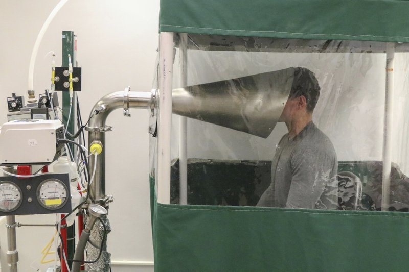 Butler said such events raise concern about aerosol spread but don’t prove it happens. There could be another way for tiny particles to spread. They may not necessarily come directly from somebody’s mouth or nose, says William Ristenpart of the University of California, Davis. His research found that if paper tissues are seeded with influenza virus and then crumpled, they give off particles that bear the virus. So people emptying a wastebasket with tissues discarded by somebody with COVID-19 should be sure to wear a mask, he said. Scientists who warn about aerosols say current recommendations still make sense. Wearing a mask is still important, and make sure it fits snugly. Keep washing those hands diligently. And again, staying farther apart is better than being closer together. Avoid crowds, especially indoors. Their main addition to recommendations is ventilation to avoid a buildup of aerosol concentration. So, the researchers say, stay out of poorly ventilated rooms. Open windows and doors. One can also use air-purifying devices or virus-inactivating ultraviolet light. nicholas.duke.edu/news/online-tool-calculates-risk-classroom-transmission-airborne-covid-19 |
|
|
|
Post by Admin on Oct 13, 2020 19:26:39 GMT
A 25-year-old was infected twice with the coronavirus earlier this year, scientists in Nevada have confirmed. It is the first confirmed case of so-called reinfection with the virus in the U.S. and the fifth confirmed reinfection case worldwide. The cases underscore the importance of social distancing and wearing masks even if you were previously infected with the virus, and they raise questions about how the human immune system reacts to the virus. The two infections in the Nevada patient occurred about six weeks apart, according to a case study published Monday in the medical journal The Lancet. The patient originally tested positive for the virus in April and had symptoms including a cough and nausea. He recovered and tested negative for the virus in May. But at the end of May, he went to an urgent care center with symptoms including fever, cough and dizziness. In early June, he tested positive again and ended up in the hospital. "The second infection was symptomatically more severe than the first," the authors of the study write. The patient survived his second bout with COVID-19. This is the second confirmed case of coronavirus reinfection in which the patient was sicker the second time. A patient in Ecuador also suffered a more serious case of COVID-19 the second time they were infected with the virus. Scientists are unsure why this might be. In theory, the body's immune system should make antibodies after the first infection that help it combat the virus more effectively if the person is exposed to the same virus again. "There are many reasons why a person might get sicker the second time around," explains Akiko Iwasaki, a professor of immunobiology at Yale University who was not involved in the Nevada study. For example, "they may have been exposed to a lot higher levels of the virus the second time around," she says, or the immune response from the first infection might be making the disease worse rather than better. But, she stresses, "this is all very speculative" because scientists still have very little information about the mechanisms at play. One of the biggest outstanding questions is how widespread reinfection might be. It's difficult to confirm cases in which a person is infected twice. Scientists must have the nasal swabs from both the first and second infection in order to compare the genomes of both virus samples.  Only the most advanced hospital and laboratory facilities have the equipment and personnel to do the genome sequencing and analyze the results. As a result, most cases of reinfection are likely going undetected. Danny Altmann, a professor of immunology at Imperial College London, says it seems that about 90% of people who have experienced "a clear, symptomatic infection" have the antibodies to fight off another infection, "perhaps for about a year." "Of course, that leaves 10% who don't" have sufficient antibodies to fight off a second infection, he wrote in an email to NPR. "[T]hey have precisely the same risk as anyone out there, thus a small but significant number of reinfections." The authors of the new study also raise the possibility that cases of people being infected multiple times could have implications for the efficacy of a coronavirus vaccine, since some people exposed to the virus may not be mounting sufficient immune responses to protect themselves from a second infection. But Iwasaki says such cases have no bearing on the efficacy of a future vaccine. The virus can deploy proteins to get in the way of the immune response, whereas a vaccine has none of those proteins, she explains. "The good thing about a vaccine is that it can induce much better immunity, a much longer lasting immunity, than the natural exposure to the the virus," she says. |
|
|
|
Post by Admin on Nov 6, 2020 4:12:23 GMT
One of the more surprising symptoms of COVID-19 has been the blood clots that many patients, including younger ones, have experienced with the infection. The clots have in some cases led to dangerous blockages in the lungs, caused strokes and even death, even in people without a history of circulatory conditions.
In a paper published in Science earlier this week, researchers provide a glimpse into what may be driving the clots triggered by COVID-19 infection. The group found that a specific set of antibodies known as autoantibodies—which are rogue versions of cells meant to defend the body from pathogens, but instead attack its own cells (in this case the body’s own blood vessel cells)—may be partly responsible for the clotting risk associated with the disease. Among 172 patients hospitalized with COVID-19, they found that half produced these autoantibodies. In addition, when the scientists injected the autoantibodies into lab mice, the animals developed blood clots.
In April, the same group of scientists reported that the inflammation associated with COVID-19 can lead to clots in small vessels in the lungs, and that these clots are mostly packed with an immune cell known as a neutrophil. In COVID-19 patients, these neutrophils can explode inside small blood vessels, creating sticky molecular traps that attract other clotting factors circulating in the blood. “Evolutionarily, we think these are meant to trap things like bacteria or viruses,” says Yogen Kanthi, an assistant professor at the University of Michigan, investigator at the National Heart, Lung and Blood Institute and one of the study’s authors. “But if [neutrophils] are over stimulated, they can also grow and cause blockages in blood vessels and drive blood clotting.” In that earlier study, Kanthi and his colleagues found that COVID-19 patients who had more of these “traps” in their blood system were more likely to have severe disease or respiratory failure.
Prothrombotic autoantibodies in serum from patients hospitalized with COVID-19
Science Translational Medicine 02 Nov 2020:
eabd3876
DOI: 10.1126/scitranslmed.abd3876
Abstract
Patients with COVID-19 are at high risk for thrombotic arterial and venous occlusions. Lung histopathology often reveals fibrin-based occlusions in the small blood vessels of patients who succumb to the disease. Antiphospholipid syndrome is an acquired and potentially life-threatening thrombophilia in which patients develop pathogenic autoantibodies targeting phospholipids and phospholipid-binding proteins (aPL antibodies). Case series have recently detected aPL antibodies in patients with COVID-19. Here, we measured eight types of aPL antibodies in serum samples from 172 patients hospitalized with COVID-19. These aPL antibodies included anticardiolipin IgG, IgM and IgA; anti-β2 glycoprotein I IgG, IgM, and IgA; and anti-phosphatidylserine/ prothrombin (aPS/PT) IgG and IgM. We detected aPS/PT IgG in 24% of serum samples, anticardiolipin IgM in 23% of samples, and aPS/PT IgM in 18% of samples. Antiphospholipid autoantibodies were present in 52% of serum samples using the manufacturer’s threshold and in 30% using a more stringent cutoff (≥40 ELISA-specific units). Higher titers of aPL antibodies were associated with neutrophil hyperactivity including the release of neutrophil extracellular traps (NETs), higher platelet counts, more severe respiratory disease, and lower clinical estimated glomerular filtration rate. Similar to IgG from patients with antiphospholipid syndrome, IgG fractions isolated from COVID-19 patients promoted NET release from neutrophils isolated from healthy individuals. Furthermore, injection of IgG purified from COVID-19 patient serum into mice accelerated venous thrombosis in two mouse models. These findings suggest that half of patients hospitalized with COVID-19 become at least transiently positive for aPL antibodies and that these autoantibodies are potentially pathogenic.
INTRODUCTION
Abnormal coagulation characteristics correlate with COVID-19 severity (1, 2). The presence of high D-dimer concentrations in plasma is an independent risk factor for death (1, 3–5). Early descriptions of COVID-19 coagulopathy identified this disorder as disseminated intravascular coagulation. However, most patients maintain normal concentrations of coagulation factors, fibrinogen, and platelets suggesting that COVID-19 induces a unique prothrombotic state that is distinct from traditional descriptions of sepsis-induced coagulopathy (6, 7). There are now increasing reports of venous thromboembolism in patients with COVID-19 (8, 9). This observation is despite concerns regarding under-diagnosis given baseline elevations in the biomarker D-dimer, as well as pragmatic challenges in obtaining diagnostic imaging while patients are in isolation. Arterial thrombosis including strokes and myocardial infarctions have also been described (9, 10). Histopathology of lung specimens from patients with severe disease shows not only characteristic findings of ARDS, but also evidence of fibrin-based occlusion of small blood vessels (11–13). There are several, possibly synergistic mechanisms by which SARS-CoV-2 infection may result in macrovascular and microvascular thrombosis (14). These include a cytokine storm that activates leukocytes, endothelium, and platelets; hypoxic vaso-occlusion; and direct activation of immune and vascular cells by virus infection. Furthermore, many patients hospitalized with COVID-19 exhibit neutrophil extracellular traps (NETs) in their blood (15, 16) and these inflammatory cell remnants may also contribute to the prothrombotic milieu (17–20).
Antiphospholipid syndrome is an acquired thrombophilia, affecting at least 1 in 2000 individuals (21). Patients form durable autoantibodies to phospholipids and phospholipid-binding proteins (aPL antibodies), such as prothrombin and beta-2-glycoprotein I (β2GPI). These autoantibodies engage cell surfaces, where they activate endothelial cells, platelets, and neutrophils (22, 23), thereby tipping the blood-endothelium interface toward thrombosis. A key feature of antiphospholipid syndrome is its ability to promote thrombosis in vascular beds of all sizes, including both arterial and venous circuits. The catastrophic variant of antiphospholipid syndrome is frequently fatal, and bears some similarities to the diffuse coagulopathy seen in patients with COVID-19 (24). Classification criteria for antiphospholipid syndrome (last updated in 2006) seek persistently positive testing for anticardiolipin autoantibodies (aCL antibodies) or anti-β2GPI autoantibodies (aβ2GPI antibodies) (25). The lupus anticoagulant test (a functional assay that screens for aPL antibodies based on their paradoxical ability to prolong in vitro clotting assays) is also included in the criteria and detects a variety of species of aPL antibodies including anti-phosphatidylserine/prothrombin autoantibodies (aPS/PT antibodies) (26).
Reports of aPL antibodies in COVID-19 and their possible relationship to thrombosis have begun to emerge in case reports and case series (27–32). Whereas viral infections are well-known triggers of transient aPL antibody production (33–36), the extent to which these short-lived autoantibodies are pathogenic has not been well defined. Here, we aimed to test for several types of aPL antibodies in serum samples from a cohort of 172 patients hospitalized with COVID-19. We also asked whether purified IgG fractions from these patients had prothrombotic properties in vitro and in two mouse models of thrombosis.
|
|
|
|
Post by Admin on Nov 6, 2020 19:27:59 GMT
RESULTS
Prevalence of aPL antibodies in serum from hospitalized COVID-19 patients
Serum samples from 172 patients hospitalized with COVID-19 (table S1) were evaluated for eight different types of aPL antibodies. Of the 172 patients, 19% died and 8% remained in the hospital at the time of this analysis. Eighty-nine patients tested positive for at least one type of aPL antibody based on the manufacturer’s cut-off, representing 52% of the entire cohort (Table 1). The lupus anticoagulant test, a functional assay that relies on altered coagulation times in plasma to detect antiphospholipid antibody activity, was not performed here given lack of access to fresh plasma samples. Among the various aPL antibodies tested, aPS/PT IgG had the highest prevalence (24%), followed by aCL IgM (23%) and aPS/PT IgM (18%) (Table 1). Forty-one patients (24%) were positive for more than one type of aPL antibody and 13 (8%) were positive for more than two types of aPL antibody. Fifty-two patients (30%) had at least one moderate- to high-titer aPL antibody (Table 1). Thirty-six patients had serum samples taken at multiple time points available for aPL antibody testing, which enabled longitudinal analysis (fig. S1). In Table 1, the highest available aPL antibody serum titer was used to classify positivity for each of these 36 patients . Seropositivity was also assessed using only the first available serum sample, with similar rates of positivity as presented in Table 1 (Table S2). To further elucidate the antigen specificity of autoantibodies in serum samples positive for aPS/PT antibodies, we measured anti-phosphatidylserine autoantibodies (aPS antibodies) in these serum samples. Neither aPS IgG nor aPS IgM correlated with aPS/PT antibody serum titers suggesting that aPS/PT antibodies in COVID-19 patient serum primarily recognized prothrombin (fig. S2). In summary, serum samples from 52% of patients hospitalized for COVID-19 were positive for aPL antibodies, with approximately two-thirds of those being detected at moderate-to-high titers. The majority of positive serum samples were associated with three types of autoantibodies: aPS/PT IgG, aCL IgM, and aPS/PT IgM.
Table 1 Prevalence of antiphospholipid antibodies in serum from COVID-19 patients (n=172)
aPL antibody Number positive (manufacturer’s cut-off) % Number positive
(titer ≥40 units) %
aCL IgG 8 4.7% 2 1.2%
aCL IgM 39 23% 13 7.6%
aCL IgA 6 3.5% 1 0.58%
aβ2GPI IgG 5 2.9% 3 1.7%
aβ2GPI IgM 9 5.2% 7 4.1%
aβ2GPI IgA 7 4.1% 3 1.7%
aPS/PT IgG 42 24% 21 12%
aPS/PT IgM 31 18% 21 12%
any positive aPL 89 52% 52 30%
aPL antibody, antiphospholipid autoantibodies; aCL, anticardiolipin antibodies; aβ2GPI, anti-beta-2 glycoprotein I antibodies; aPS/PT, anti-phosphatidylserine/prothrombin antibodies;
Manufacturer’s cut-off: aCL IgG, IgM, IgA = 20 IgG, IgM, IgA phospholipid units; aβ2GPI IgG, IgM, IgA = 20 standard IgG, IgM, IgA units; aPS/PT IgG, IgM = 30 units
Clinical correlates of aPL antibodies
We next asked whether the presence of aPL antibodies was associated with various clinical characteristics. Specifically, we assessed potential correlations of aPL antibodies with the ratio of oxygen saturation to fraction of inspired oxygen (SpO2/FiO2, i.e., oxygenation efficiency), C-reactive protein in serum, D-dimer concentrations in plasma, platelet counts, absolute neutrophil counts, calprotectin in serum (a marker of neutrophil activation), and myeloperoxidase (MPO)-DNA complexes in serum (markers of neutrophil extracellular traps, NETs) (Table 2). Titers of aCL IgM correlated with all of these clinical and laboratory variables (Table 2). Neutrophil activation as indicated by calprotectin in serum was most consistently associated with the presence of aPL antibodies (Table 2). We also assessed a previously devised tool called the aPL score, which integrates and prioritizes data from the various aPL antibody types tested (37). The aPL score demonstrated a positive correlation with platelet count (p=0.025), neutrophil activation (p=0.0007), and the presence of NETs (p=0.02) (Table 2).
Table 2 Correlation of antiphospholipid antibodies with clinical and laboratory variables in COVID-19 patients‡
aPL Score
(modified) aCL IgG aCL IgM aβ2GPI IgG aβ2GPI IgM aPS/PT IgG aPS/PT IgM
Spearman r p r p r p r p r p r p r p
Clinical and lab variables
SpO2/FiO2 -0.051 ns -0.16 * -0.19 * -0.10 ns -0.022 ns -0.11 ns -0.16 *
C-reactive protein 0.031 ns 0.15 ns 0.17 * 0.075 ns -0.040 ns 0.058 ns 0.16 *
D-dimer 0.087 ns 0.092 ns 0.24 ** 0.041 ns 0.000 ns 0.005 ns 0.037 ns
Platelet count 0.17 * 0.095 ns 0.29 **** 0.17 * 0.11 ns -0.009 ns 0.23 **
Neutrophil count 0.10 ns 0.13 ns 0.19 * 0.047 ns 0.041 ns -0.008 ns 0.096 ns
Calprotectin 0.26 *** 0.29 **** 0.28 *** 0.11 ns 0.090 ns 0.25 *** 0.23 **
NETs (MPO/DNA) 0.18 * 0.16 * 0.25 *** 0.20 ** 0.13 ns 0.033 ns 0.23 **
ns, not significant; NETs, neutrophil extracellular traps; MPO, myeloperoxidase; *p<0.05, **p<0.01, ***p<0.001, and **** p<0.0001
|
|
|
|
Post by Admin on Nov 6, 2020 20:44:48 GMT
We then examined clinical variables as they related to positive aPL antibody thresholds for each ELISA test. A positive test for any aPL antibody was associated with higher calprotectin in serum (p=0.009) and lower clinical estimated glomerular filtration rate (eGFR, p=0.03) (Fig. 1A, B). These associations were also observed when comparing patient serum samples that were positive for aPS/PT antibodies to serum samples from the remainder of the cohort (calprotectin p=0.0008; eGFR p=0.008) (Fig. 1C,D) or serum samples without aPL antibodies (calprotectin p=0.001; eGFR p=0.01) (fig. S3). Nadir eGFR was lower in patients with a history of renal disease compared to those without (p=0.01) (fig. S4). Oxygenation efficiency tended to be impaired in patients with serum samples positive for aPL or aPS/PT antibodies compared to those whose serum samples lacked these antibodies, although group comparisons did not reach statistical significance (fig. S5). Similarly, peak troponin in serum and peak D-dimer in plasma tended to be higher in patients with a positive test for any aPL antibody or anti-PS/PT antibody, respectively (fig. S6). Given that obesity can affect the D-dimer concentration in plasma, we compared D-dimer plasma concentrations in COVID-19 patients with or without obesity, but did not find a difference (fig. S7). Thus, the presence of aPL antibodies in serum samples from patients with COVID-19 correlated with various clinical characteristics, especially neutrophil activation and impaired renal function.  Fig. 1 aPL antibodies, NET release, and renal function. Serum samples were obtained from 172 patients hospitalized with COVID-19. (A, B) Patients were divided into two groups based on whether their serum samples were positive (+) or negative (-) for the presence of aPL antibodies (positivity was based on the manufacturer’s threshold). Shown is the amount of calprotectin in serum, a measure of neutrophil activation (A), and the clinical estimated glomerular filtration rate (eGFR) (B) for the two groups. (C, D) Patients were divided into two groups based on whether their serum samples were positive (+) or negative (-) for the presence of aPS/PT antibodies (IgG and IgM considered together); manufacturer’s thresholds were used to determine positivity. Shown is the amount of calprotectin (C) and the eGFR (D) for the two groups. Groups were analyzed by an unpaired t test: *p<0.05, **p<0.01, and ***p<0.001. Horizontal black bars represent the mean. For patients who had serum samples available at multiple time points, only the first-available serum sample was used in this analysis. IgG isolated from COVID-19 patient serum triggers release of NETs Work by our group and others has revealed that one prothrombotic function of aPL antibodies in patients with antiphospholipid syndrome is to trigger release of NETs (23, 38). Given that we recently detected elevated NETs in serum from patients with COVID-19 (15), we reasoned that IgG fractions purified from serum of patients with COVID-19 might be able to trigger NET release. We selected two COVID-19 patients with high serum aβ2GPI IgG, two COVID-19 patients with high serum aPS/PT IgG, and two COVID-19 patients who lacked serum aPL antibodies. From these patients, we purified total IgG fractions and tested them alongside IgG pooled from two patients with active catastrophic antiphospholipid syndrome as well as a separate IgG pool from five patients with antiphospholipid syndrome who tested positive for aCL antibodies, aβ2GPI antibodies, and lupus anticoagulant. The purity of isolated COVID-19 patient IgG was verified by SDS-PAGE (fig. S8). To quantify NET release in vitro, we measured MPO activity released into the supernatant after digestion of NET DNA with micrococcal nuclease. The release of NETs from neutrophils isolated from healthy individuals doubled (compared with unstimulated neutrophils) when neutrophils were cultured with COVID-19 patient IgG samples positive for aPL antibodies (Fig. 2A, data file S1). This was similar to the degree of NET release induced in neutrophils by IgG samples from patients with antiphospholipid syndrome (p<0.0001) or catastrophic antiphospholipid syndrome (p=0.0001). Representative images of NET release induced by COVID-19 patient IgG are shown in Fig. 2B. We have previously shown that dipyridamole—an antithrombotic medication—can attenuate aPL antibody-mediated prothrombotic NET release by surface adenosine A2A receptor agonism (39). Here, we found that dipyridamole also suppressed COVID-19 patient IgG-mediated NET release from neutrophils in vitro (fig. S9). IgG fractions purified from COVID-19 patient serum positive for aPL antibodies promoted NET release similar to IgG isolated from individuals with established antiphospholipid syndrome.  Fig. 2 COVID-19 patient IgG promotes NET release from normal neutrophils in vitro. (A) Control neutrophils were isolated from healthy individuals and cultured in the presence of human IgG (10 μg/ml) for 3 hours. IgG fractions were obtained from COVID-19 patients who were or were not positive for aPL antibodies (aPS/PT or aβ2GPI as indicated), and from patients with antiphospholipid syndrome (APS) or catastrophic antiphospholipid syndrome (CAPS). NET release was measured by the enzymatic activity of myeloperoxidase (MPO) after solubilization of NETs with micrococcal nuclease; fold increase is plotted relative to unstimulated neutrophils. Data are derived from four independent experiments. Comparisons were by one-way ANOVA with correction for multiple comparisons by Dunnett’s method: *p<0.05, **p<0.01, ***p<0.001. (B) Representative images show released NETs, indicated by yellow arrows. DNA, blue; neutrophil elastase, green. Scale bar,100 microns. IgG isolated from aPL antibody-positive patient serum potentiates thrombosis in mice We next sought to determine whether IgG fractions from COVID-19 patient serum could accelerate thrombosis. When tested in an in vitro cell-free thrombin generation assay, IgG fractions purified from COVID-19 patient serum did not have demonstrable clot-accelerating activity (fig. S10). Nevertheless, we speculated that a prothrombotic phenotype might still be observed in the cell-enriched vascular environment of mice. We have previously reported that IgG isolated from serum of patients with either triple-positive antiphospholipid syndrome or catastrophic antiphospholipid syndrome accelerates large-vein thrombosis in various mouse models of inferior vena cava thrombosis (38–40). Here, we asked whether COVID-19 patient serum IgG might behave similarly to enhance thrombosis in these mouse models. We first used a mouse model in which a copper wire was placed inside the inferior vena cava in order to activate the endothelium by electrolysis-mediated free radical generation (Fig. 3A). In this model, IgG isolated from COVID-19 patients with a high serum titer of aPS/PT IgG increased thrombus extension (Fig. 3B) and overall accretion (Fig. 3C,D; data file S2) 24 hours after IgG intravenous injection. The high aPS/PT serum titer samples also increased NET remnants in mouse serum (p=0.0004), similar to IgG from patients with catastrophic antiphospholipid syndrome (p=0.0014) (Fig. 3E; data file S3), and demonstrated a tendency toward higher expression of citrullinated histone H3 (a biochemical marker of NETs) in mouse thrombi by Western blotting (fig. S11). To confirm these findings, we turned our attention to an independent mouse model in which the inferior vena cava was narrowed just distal to the renal vein by a fixed suture placed over a spacer that was subsequently removed (Fig. 3F); thrombus size was measured 24 hours after IgG intravenous injection. In this “stenosis” mouse model of thrombosis, IgG from COVID-19 patients with a high aPS/PT serum titer also increased thrombus extension (p=0.01) (Fig. 3G), thrombus accretion (p=0.003) (Fig. 3H, I; data file S4), and circulating NET remnants (p=0.008) (Fig. 3J; data file S3) 24 hours after IgG intravenous injection. Taken together, these data indicate that IgG fractions from some patients with acute COVID-19 were able to accelerate thrombosis in vivo. |
|

