|
|
Post by Admin on Dec 10, 2020 19:47:42 GMT
One of the best preserved Neanderthal skeletons ever discovered apparently had wicked nasty dental health. Scientists are calling the specimen Altamura Man, named for the town in southern Italy where he fell into a hole and starved to death more than 130,000 years ago. Cavers first found his weary bones in 1993, but new revelations about his grungy choppers have scientists reaching for their notepads. Also their Colgate and dental floss, probably. Altamura Man remains lodged in the earth, and he’s only accessible about 20 minutes through the surface through a system of narrow crevices. But that didn’t stop researcher Jacopo Moggi-Cecchi and his team from snapping photos, videoscope footage, and X-rays in the cave’s silent depths. The team’s new research was published in the journal PLOS earlier this month, and it’s largely focused on the doomed Neanderthal’s teeth.  The research includes a thorough study of the man’s jaw, including his almost complete set of admittedly grimy teeth. According to Moggi-Cecchi, scientists were surprised to find that Altamura Man had been skimping on the primitive White Strips. That is, he had worse-than-usual dental health. “We have a large fossil record of Neanderthals, and it’s not typical,” Moggi-Cecchi told CNN. “In terms of oral health, they were in good shape.” Moggi-Cecchi added that the roots of some of Altamura Man’s teeth were exposed, potentially suggesting gum disease, and some teeth in the lower jaw also had significant plaque deposits. Finally, Moggi-Cecchi pointed out that, like other Neanderthals, this ancient man’s front teeth are larger than those of modern humans. Listen up, people, because I’m about to rock your world: according to Moggi-Cecchi, early man used those big-ass buck teeth as a “third hand” to hold meat while cutting it. We’ve clearly evolved—I mean, the mere sight of a spork would likely be enough to send Altamura Man into cardiac arrest. But I’m currently picturing Altamura Man using his buck tooth to saw away at a piece of woolly rhinoceros, and I can’t help but ask Mother Nature: why’d you have to go and mess with perfection? |
|
|
|
Post by Admin on Dec 11, 2020 19:37:10 GMT
Altamura Man is one of the most complete and best preserved Neanderthal skeletons ever discovered. His fossilized bones, however, have remained hidden from view at the bottom of a sinkhole near Altamura, a town in southern Italy. That's where he fell and starved to death more than 130,000 years ago. 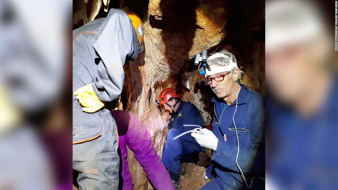 Cavers came face to face with his skull, covered in limestone deposits, for the first time in 1993. Frustratingly for scientists, though, its inaccessible location -- a 20-minute journey from the surface through narrow crevices -- has made study of the skeleton extremely difficult. The body remains lodged in a small chamber deep in the karst cave system. "They used the rope to bring me down and many of my colleagues. For me, it was a totally amazing experience. When you get in that corner and you see the skeleton there, you're really blown away," said Jacopo Moggi-Cecchi, a professor in the department of biology at the University of Florence. "This individual must have fallen down a shaft. Maybe he didn't see the hole in the ground. We think he sat there and died," said Moggi-Cecchi. "The original shaft he fell through is no longer there. It's been filled by sediment so we are confident the entire skeleton is there. No animals could have got there." 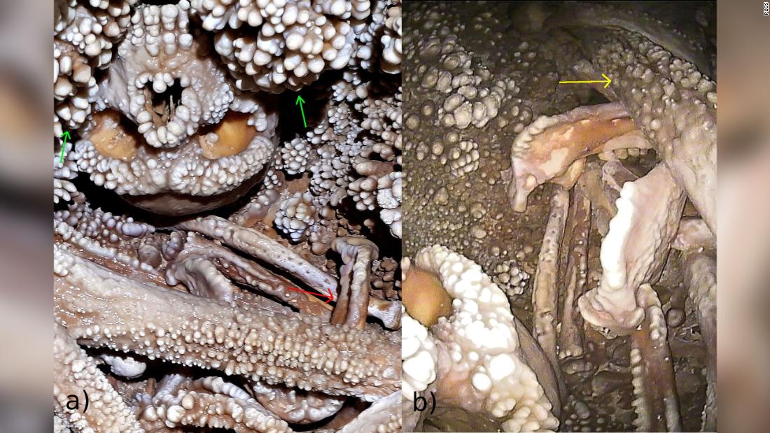 This new research, published in the journal PLOS on Wednesday by Moggi-Cecchi and his colleagues, is beginning to yield more information about the man. Based on photos, videoscope footage and X-rays taken in the depth of cave, scientists have published an initial study of the man's jaw, including an almost complete set of teeth. They suggest that the man was of adult age, but not old, and he had also lost two teeth before he died. "The tooth loss is something interesting. We have a large fossil record of Neanderthals, and it's not typical. In terms of oral health, they were in good shape," said Moggi-Cecchi. The roots of some teeth were exposed, which could suggest gum disease was at play, he said. Some teeth in the lower jaw also had deposits of dental calculus -- calcified plaque that's familiar to dentists today. Homo neanderthalensis walked the Earth for a period of about 350,000 years before they disappeared, living in what's now Europe and parts of Asia. They disappeared about 40,000 years ago -- although it's believed that they overlapped with Homo sapiens geographically for a period of more than 30,000 years after some humans migrated out of Africa. Earlier research, published in 2016 based on DNA analysis of the man's shoulder bone, confirmed that the body was indeed Neanderthal and that he had lived between 130,000 to 172,000 years ago.Our archaic relatives used their front teeth almost as a "third hand" to hold meat while cutting it or to hold skins or leather for preparation, Moggi-Cecchi explained. Altamura Man had "marked wear" that might be related to this kind of activity.  For a more detailed analysis, however, Moggi-Cecchi said that it would be necessary to get the skull inside a lab as the teeth, like the rest of the skeleton, are covered in calcite -- mineral deposits from the limestone karst. Scientists hope one day that the skeleton, or at least part of it, will be removed from the cave to allow in-depth study. Ultimately, Moggi-Cecchi said the man could become a Neanderthal version of Otzi the Iceman -- whose 5,300-year-old frozen body was found by a couple hiking in the North Italian Alps in 1991. Otzi has become a window into early human history for scientists and tourists alike. Nearly every part of him has been analyzed, including what he may have sounded like, the contents in his stomach and how he died. "The fact that we can get this kind of information simply by looking at the specimen in situ, imagine what the possibilities are if we can extract the specimen from the cave. " |
|
|
|
Post by Admin on Dec 12, 2020 20:43:38 GMT
In situ observations on the dentition and oral cavity of the Neanderthal skeleton from Altamura (Italy) Alessandro Riga,Marco Boggioni,Andrea Papini,Costantino Buzi,Antonio Profico,Fabio Di Vincenzo,Damiano Marchi,Jacopo Moggi-Cecchi ,Giorgio Manzi Published: December 2, 2020 doi.org/10.1371/journal.pone.0241713Abstract The Neanderthal specimen from Lamalunga Cave, near Altamura (Apulia, Italy), was discovered during a speleological survey in 1993. The specimen is one of the most complete fossil hominins in Europe and its state of preservation is exceptional, although it is stuck in calcareous concretions and the bones are mostly covered by calcite depositions. Nevertheless, it is possible to carry out some observations on craniodental features that have not previously been described. In this work, we present an account of the oral cavity, made possible by the use of a videoscope, which allowed us to reach some hidden parts of the mandible and palate. This is the first detailed overview of the teeth and maxillary bones of the Neanderthal skeleton from Altamura. The dentition is almost complete. However, two teeth (upper right P3 and upper left M1) were lost ante mortem and four teeth (lower right I1 and P3 and lower left I1 and I2) were lost most probably post mortem. Dental wear is marked. The erupted M3s and the inversion of the compensating curve of Wilson in the M1s and M2s but not in the M3s suggest that the individual is fully adult, but not old. Although most of the teeth have their roots exposed for several millimeters, the periodontal bone appears to be in good condition overall, except in correspondence of the two ante-mortem tooth losses. X-rays of the anterior teeth show a periapical lesion, probably linked to the advanced dental wear. We also observed a weak expression of taurodontism in the posterior dentition and the presence of a retromolar space, features consistent with an attribution to the Neanderthal hypodigm; this attribution is also supported by aspects of the cranial morphology, the morphometric analysis of the scapula and preliminary mtDNA data. There is also a well-developed palatine torus, to the best of our knowledge a feature not previously described in Neanderthals. Introduction The Neanderthal skeleton from Altamura (Apulia, Italy), discovered in 1993 [1], is the most complete Neanderthal ever found and possibly the most complete fossil hominin ever found before modern humans. It was discovered in a branch of Lamalunga Cave (40° 52' 18.64" N, 16° 35' 14.98" E), which is the upper part of a wider karstic complex in the Murgia plateau. This geographic region of southern Italy is rich in karstic formations such as dolines, caves and blind valleys [2]. The skeleton lies in a corner of a small chamber (Fig 1), called the “Apse of Man”, delimited by speleothemic formations and embedded in calcareous formations, with all the bones covered by a calcite layer and coralloid deposits [1, 3]. Faunal elements found in the cave were probably scattered by water, whereas all the bones of the hominin specimen are concentrated in a small area and it is likely that the skeleton now lies approximately where the body decomposed [4, 5]  Fig 1. The “Apse of Man”. The Altamura specimen lies in a corner of this small chamber, mostly embedded in calcareous formations and covered by calcite layers and coralloid deposits. Observations of the skeletal features suggest a combination of archaic and Neanderthal derived traits, placing the specimen in the human variability of the Late-Middle Pleistocene in Europe [6]. Between 2009 and 2015, a large part of its fragmentary right scapula was removed from the cave in three pieces [3, 7, 8]. In particular, the portion comprising the glenoid fossa, the neck and the roots of the spine and coracoid process of the scapula was extracted in 2009 and its study provided information on paleogenetics and morphology [3]. The morphometric analysis of the glenoid fossa confirmed that the specimen falls within the Neanderthal range of variation, despite some morphological peculiarities [3, 8]. At the same time, paleogenetic data confirmed the Neanderthal attribution [3] of the specimen, while U/Th dating suggested that the individual died between 130.1 ± 1.9 ka and 172 ± 15 ka [3]. Within the context of a broader project coordinated by one of us (GM) and financed by the Italian Ministry of Instruction, University and Research (MIUR; PRIN call 2015), new research activities were recently carried out in the cave. During this new stage, high-definition photographic and three-dimensional (3D) digital images were acquired by different techniques, allowing the observation of several new features of the Altamura Neanderthal despite the presence of the calcareous concretions partially covering the specimen. In this work, we present the first observations carried out in situ on the dentition and oral cavity. |
|
|
|
Post by Admin on Dec 12, 2020 23:42:32 GMT
Materials and methods To understand what kind of observations would be possible, it was important to figure out the relative position of the cranium and mandible in the cave (Fig 2). The cranium lies on its vault, slightly tilted to the left, with the orbits facing anteriorly toward the ‘Apse’. Two large apices of karstic concretions descend from above, framing the midface on the right and the left side of the cranium, thus preventing direct observation of the entire maxillary arcade and teeth. Only the labial aspect of the incisors can be easily examined. The mandible is located in the bone assemblage in front of the cranium and slightly to its right (Fig 2). It lies upside down with the teeth facing the floor of the cave. The occlusal surfaces of the molar teeth are in partial contact with the right fibula below them. The right femur rests above the anterior part of the body of the mandible, close to the symphysis.  Fig 2. Cranium and mandible. a) the cranium lies on its vault, embedded between two large calcareous formations (green arrows), while the mandible is in front of the cranium, slightly to its right (red arrow). b) The mandible lies upside down above other bones and the right femur (blue arrow) rests on it, covering the anterior part of the body and the mandibular symphysis. Overall preservation of the dentition and oral structures is good; however, all surfaces (teeth and bones) are covered with a calcite layer of variable thickness. Moreover, large “coralloid” formations (as defined in [3]) cover the tooth crowns in some areas, notably the maxillary anterior teeth, partially hiding the original morphology of the labial, incisal and lingual surfaces (Fig 2A). Because of the position of the cranium and mandible and the widespread calcite layers covering all bones and teeth, morphological observations were limited to the gross morphology of the oral structures, and any analysis had to be carried out with the help of various technical devices. Photos of the dentition, maxilla and mandible were taken with an Olympus IPLEX NX IV9635N videoscope with a 3.5 m-long probe of 6 mm diameter and 120D/NF lens with a 120° angle; the base unit was an 8.4-inch daylight view LCD touchscreen monitor. The small dimensions and the high flexibility of the Olympus probe meant that it could be inserted through the narrow passages of the karstic flow above the cranium to provide images of the lingual side and occlusal plan of the maxillary arch and to reach the mandible below the bone assemblage. Prior to the use of the Olympus videoscope, test acquisitions were made with an Ambu aScope 3 videoscope with aView LCD monitor. The position of the skull and mandible prevented us from using X-ray devices on all the dentition and we could only take X-ray images of the upper anterior teeth with a handheld KaVo NOMAD™ Pro 2 X-ray system (exposure time 1 sec, 60 kV, 2.5 mA, distance from subject 35 cm). Phosphorus sensors were developed using a KaVo ScanXam™ scanner. Results The dentition of the Altamura Neanderthal is 80% complete; all the teeth are present in situ except for the right P3 and left M1 in the maxilla and the right I1 and P3 and left I1 and I2 in the mandible. Observations were made on the following features: ante-mortem tooth loss, periodontal status, dental wear, palatine torus, taurodontism, retromolar space and periapical lesion of the upper I2. Ante-mortem tooth loss The position of the cranium allows us to assume that the two missing maxillary teeth (RP3 and LM1) were lost in vitam. In the place for the RP3 (Fig 3A), the remnants of the alveolus are not resorbed and a deep gap is evident in the alveolar bone; the space left in the tooth row by the tooth loss is reduced due to migration of the adjacent P4.  Fig 3. Ante-Mortem Tooth Loss (AMTL). a) Maxillary right hemiarch with (from above): M1, P4, C’, I2; between P4 and C’ there is a space with the remnants of the socket of RP3; the space left in the tooth row by the tooth loss is reduced by the relative drift of adjacent teeth. b) Socket of LM1 with a well-defined space for three roots; there are no evident marks of bone resorption. We cannot exclude the possibility of a post-mortem tooth loss, but the position of the skull makes this hypothesis unlikely. The socket of LM1 is evident, although a calcareous film over the bone makes its observation difficult. The socket shows a well-defined trifurcation of the space for the roots (Fig 3B). In the mandibular dentition, the calcite deposits prevent observation of the alveolar bone and make it difficult to reconstruct whether tooth loss of the anterior teeth occurred in vitam (as a consequence of alveolar resorption) or post mortem.  AMTL has been observed in a few Neanderthal specimens (Table 1). In one case (Gibraltar 2), the specimen is a child about 5 years old [9]; all the other specimens are advanced in age. Literature reports are not in agreement on the exact number of AMTL [10, 11]; however, in 2 of the 5 specimens (Guattari 1 and La Chapelle-aux-Saints 1), AMTL occurred in at least 25% of the observable alveoli. |
|
|
|
Post by Admin on Dec 13, 2020 21:12:18 GMT
Periodontal status Periodontium, the set of tissues that support the tooth in the jaw, is composed of alveolar bone, periodontal ligament, gingiva and cementum [12]. Pathological inflammation of the periodontium results in periodontal disease, a condition that may affect soft tissues (e.g. gingivitis) or also the alveolar bone, causing its resorption [13]. In archaeological and paleo-anthropological materials, periodontal disease is detectable only when it affected alveolar bone. In the Altamura specimen, the marks of periodontal disease are present in correspondence of the lost RP3. Alveolar resorption is advanced and a deep lesion with remodeling of the alveolar margin is still present (Fig 3A). In correspondence of the other lost tooth (LM1), there is an incipient resorption of the buccal alveolar margin (Fig 3B). In both cases, the periodontal condition is localized and linked to the events that led to loss of the teeth. On the remaining portions of the dental arcades, the calcite layer prevents observation of the morphological markers of periodontal disease visible on the bone, such as foramina, grooves, depressions or pockets. However, the general appearance of the alveolar margins does not appear to be consistent with severe periodontal disease. Nevertheless, there is evidence of marked root exposure, with a distance between the alveolar margin and the cemento-enamel junction (CEJ) sometimes of several millimeters (Fig 4). In the anthropological literature, root exposure is linked to two different mechanisms: alveolar bone resorption (due to periodontal disease) and compensative eruption [14, 15]. Several methods have been developed to distinguish between the causes [15–17]; these methods base the diagnosis of periodontal disease on the texture of the alveolar bone, assuming the healthy bone to be smooth and with no (or a few) interruptions by foramina, depressions and grooves. Until the calcite layer covering the bone can be virtually removed, it will be impossible to determine the state of the alveolar margin and the reason for the large extent of root exposure.  Fig 4. Root exposure on lower left molars. Left ramus of the mandible with (from the left): M2, M1, P4, P3, C,. The calcite layer covering teeth and bone prevents observation of the morphology of the alveolar bone. Despite the absence of clear marks of periodontal disease, there is evidence of root exposure of several millimeters. In some teeth, especially in the mandible, deposits of dental calculus are present under the calcite layer (Fig 5). Dental calculus is mineralized bacterial plaque and occurs on the crown or the root surface [12, 18]; in the living, it may irritate the gums and, together with dental plaque, is among the main causes of gingivitis and periodontal disease [19]. In this specimen, observations suggest that calculus deposits are just below the CEJ, above the edge of the alveolar bone and above the root furcation. This condition suggests that the periodontal tissue near the bone was healthy, with no evidence of any active periodontal disease.  Fig 5. Dental calculus and taurodontism. On the lower right molars, as well on other teeth, dental calculus is present below the CEJ (red arrow). The position of the root furcation in RM1 is at about half of the tooth height and can thus be classified as hypotaurodont, following [20]. |
|

